Microscope dentistry: why I treat under magnification
Root canals are among the smallest, most intricate spaces in the body. I carry out every case under a dental operating microscope, because seeing that detail clearly is what makes modern endodontic treatment so much more predictable.
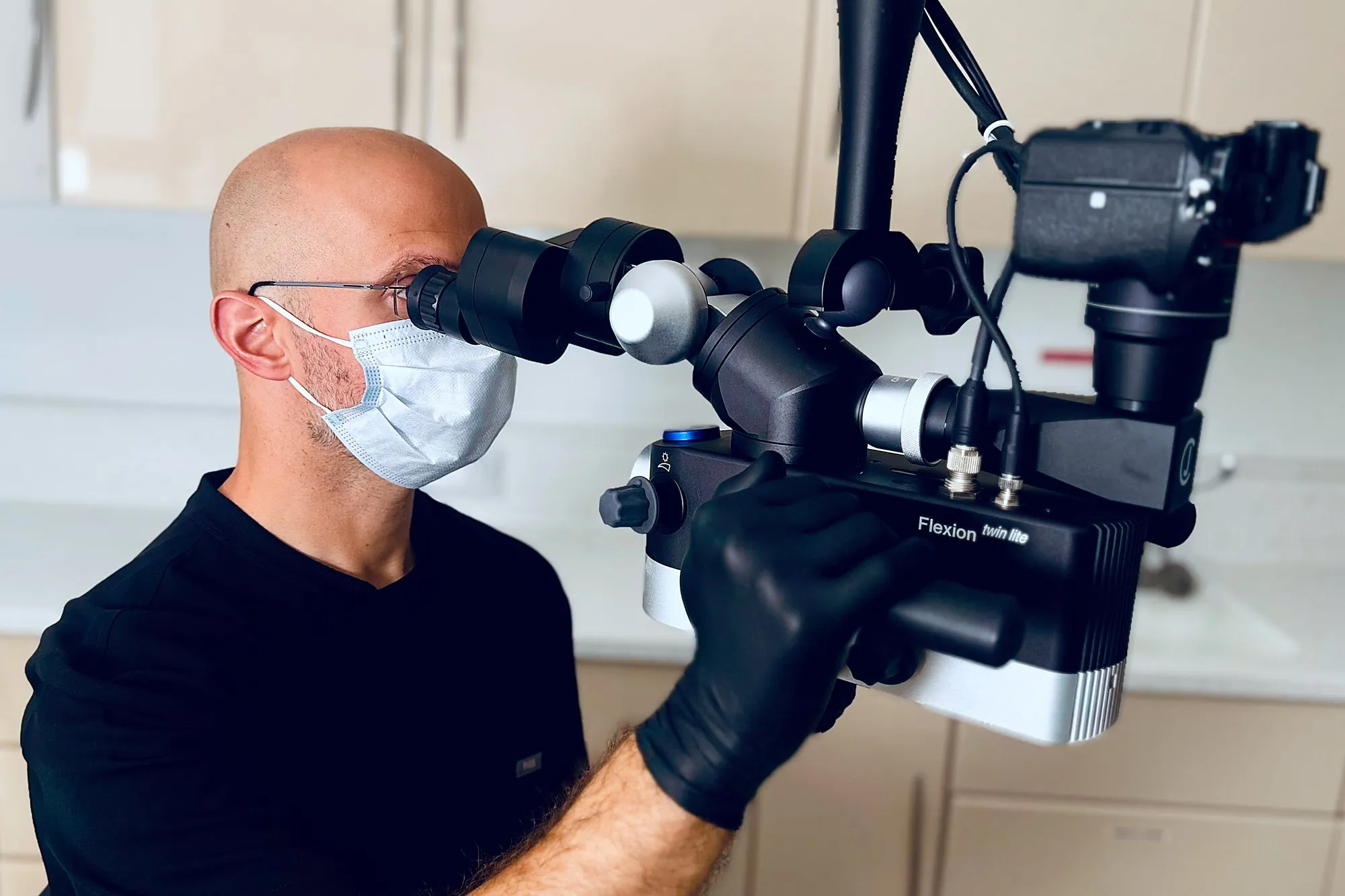
At the dental operating microscope during treatment.
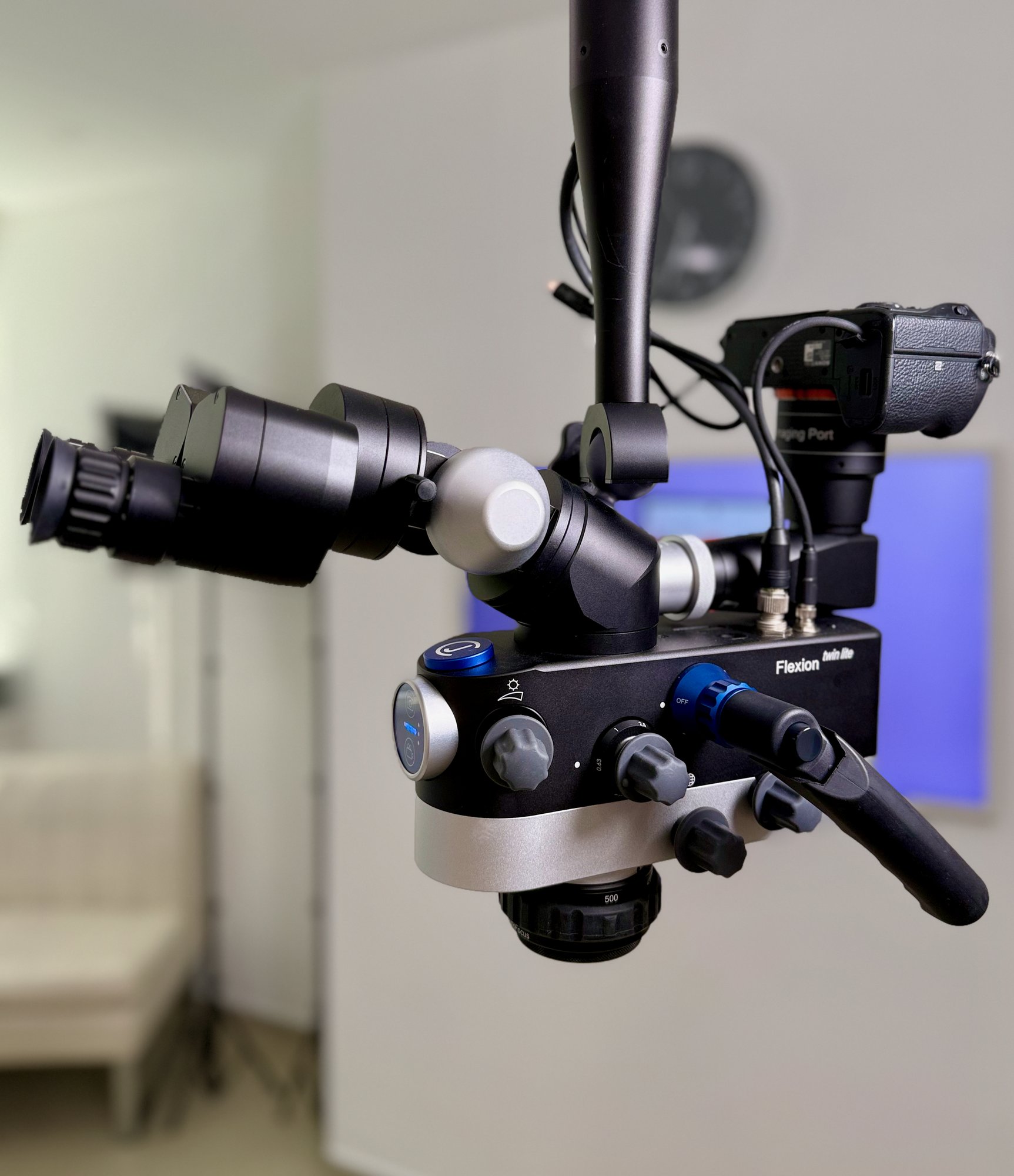
My dental operating microscope - bringing the fine detail of each root canal into clear, magnified view.
What is a dental operating microscope?
The dental operating microscope is a high-powered instrument with dedicated lighting, positioned over the tooth, that magnifies the area many times over, giving an ultra-clear, high-definition view. Magnification is the same principle surgeons use for delicate work - bringing tiny structures into sharp focus.
Why magnification matters
Root canals are fractions of a millimetre wide, often curved, and frequently include extra canals that are easy to miss. Under magnification I can find them, clean them thoroughly, and see fine cracks or detail that simply aren’t visible to the naked eye.
Preserving your natural tooth
Seeing clearly means I can be precise - removing infection while conserving as much healthy tooth as possible. Magnification is often what makes saving the tooth possible.
What magnification means for you
More accurate treatment, a higher chance of success, and often the whole thing completed in a single visit.
Seeing what I see
Because I’m working under the dental operating microscope, I can also take high-magnification photographs of your tooth during treatment, and show you exactly what I’m seeing - a hidden crack, an extra canal, or the tooth before and after - making it far easier to explain what’s going on and why a particular approach makes sense. Those photographs turn a procedure that’s hard to picture into something we can look at and talk through together.
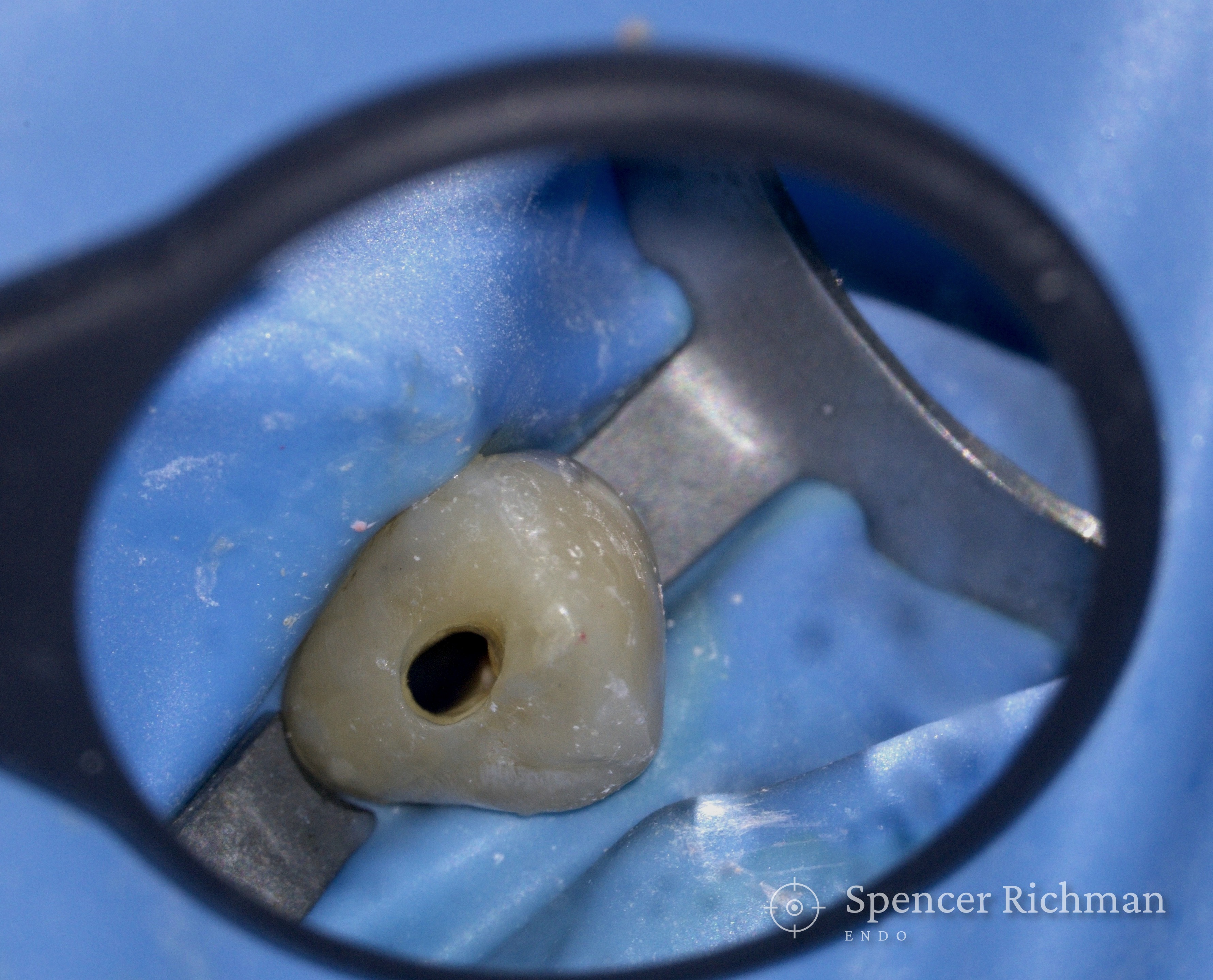
During re-treatment: the canal located and cleaned, the upper canine isolated under rubber dam and seen through the dental operating microscope.

After re-treatment: the same canal cleaned and filled.
Is the dental operating microscope standard everywhere?
Not every clinician or practice works this way. Treating every case under the dental operating microscope as standard is central to how I practise.
The difference magnification makes
The dental operating microscope isn’t technology for its own sake - the instrument is an essential piece of equipment that lets me give you an honest assessment and the best possible chance of keeping your tooth.
Get in touch