An upper molar with a missed canal: root canal re-treatment thirteen years on
An upper first molar, root treated thirteen years earlier, that had become tender to bite. A CBCT scan showed a canal that had never been found.
Presentation
A woman in her late thirties was referred by her own dentist with discomfort and tenderness around an upper left molar. The tooth had been root treated about thirteen years earlier.
The tooth was tender to percussion. Pressing on the gum over the mesio-buccal root was tender too, which points at the end of that particular root rather than at the tooth as a whole.
Assessment
The periapical X-ray showed apical pathology on the mesio-buccal root.
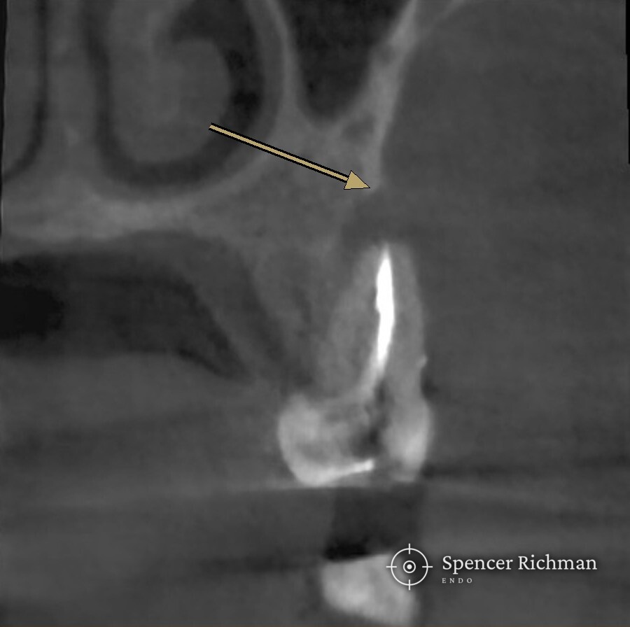
The CBCT scan showed the size and extent of the lesion, and the reason behind the lesion: a canal in the mesio-buccal root that had never been treated.
Most upper first molars carry a second canal in the mesio-buccal root, known as the MB2. The MB2 sits behind the main mesio-buccal canal, often under a shelf of dentine, and is narrow enough to be missed without magnification. Of all the canals in the mouth, the MB2 is the one most often left untreated.
An untreated canal keeps its original contents. Bacteria in that space sit beyond the reach of anything placed in the canals around them, which is how a tooth stays comfortable for years and then turns tender.
Diagnosis: chronic apical periodontitis, now symptomatic, in a previously root treated tooth, caused by anatomy that had not been treated.
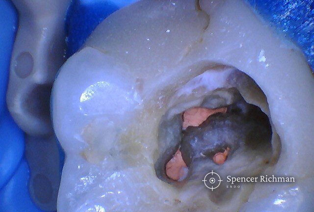
The tooth at the start: old root filling material visible in the canal openings.
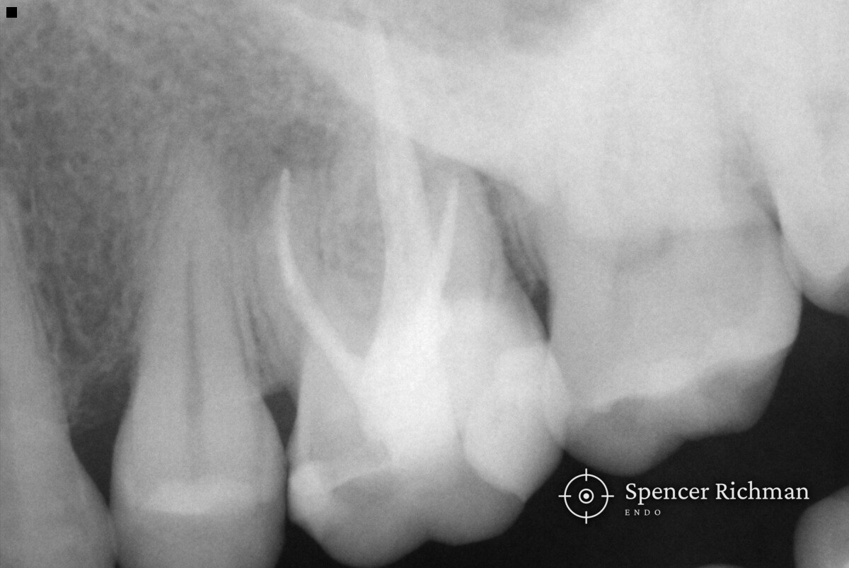
Before treatment
Treatment
Treatment was carried out over two visits at my Woodford Green practice.
Opening the tooth showed how the original filling had been placed. A carrier-based technique had been used, which leaves a plastic carrier running down the length of each canal, coated in filling material.
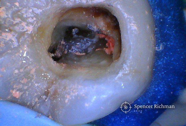
Removing a carrier is slower than removing a conventional root filling. Nothing dissolves the plastic, so each carrier has to be freed from the canal wall and drawn out whole.
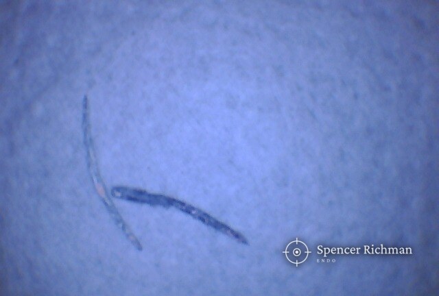
Behind the main mesio-buccal canal, the MB2 was found and treated.
The amount of work in clearing the old material made two visits the sensible plan. The tooth was dressed in between, and her symptoms had gone by the time she returned.
The second visit completed the filling, the MB2 included. She has gone back to her own dentist for an onlay, since a root treated molar has lost structure and cuspal protection stops the tooth breaking.
Why a treated tooth sometimes needs doing again is covered in the guide on root canal re-treatment.
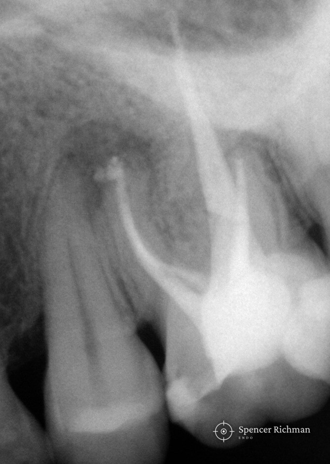
After treatment
Review
Questions patients ask
Why has my root canal started hurting years later?
An untreated canal keeps its original contents, sealed off from the rest of the tooth. That space can stay quiet for years before the response at the end of the root builds to the point where biting becomes tender.
How is a canal missed during root canal treatment?
The MB2 is narrow, lies directly behind the main mesio-buccal canal, and is often covered by a shelf of dentine. Finding the MB2 reliably needs magnification and a deliberate search for that canal.
Why do I need a CBCT scan when I have already had an X-ray?
A flat X-ray compresses a three-dimensional tooth into a single plane, so a canal sitting directly behind another stays hidden. A CBCT scan separates the two and shows how far the change in the bone extends.
Why does root canal re-treatment take two visits?
Clearing an old filling of this kind is slow work, and the tooth had infection at the end of a root. Two visits allowed the old material to come out properly and the tooth to settle before the new filling went in.
Why does a root treated molar need an onlay?
Back teeth take heavy biting loads, and a root treated molar has lost structure. An onlay covers the cusps and holds the tooth together.
Published with the patient’s written consent and fully anonymised. Every tooth is different and outcomes vary from case to case, so this case should not be read as a promise of a particular result.
Get in touch