An upper front tooth: external cervical resorption after a teenage injury
A man in his forties with no pain at all, who noticed a lump in his gum above an upper front tooth. The cause traced back to a knock taken as a teenager.
Presentation
A man in his forties came with no symptoms whatsoever. What brought him in was a lump he had noticed in the gum above an upper front tooth.
The tooth was comfortable and had never given him trouble. He did remember taking an elbow to the face during a football match as a teenager, and that knock had landed on this tooth.
Assessment
The upper left central incisor gave no response to electric pulp testing, and none to Endofrost. A tooth silent to both tests has lost the nerve supply inside.
The lump turned out to be a sinus tract, draining infection from within the tooth.
The X-ray showed a round, well-defined area of loss at the neck of the tooth, and a larger area of infection around the root end.
A CBCT scan showed the shape and depth of the defect: external cervical resorption on the palatal surface, eating into the tooth from the outside.
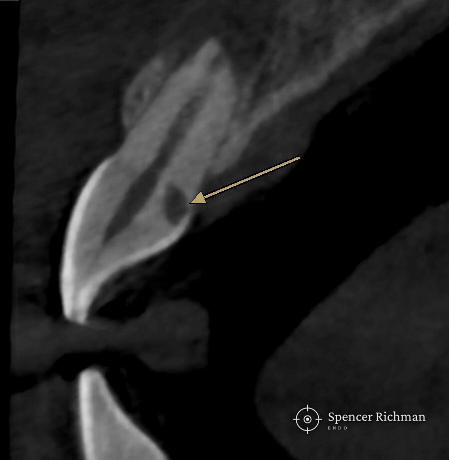
Diagnosis: external cervical resorption in a non-vital upper central incisor, with an associated sinus tract.
Resorption of this kind often follows an injury years earlier. Cells that normally remodel bone begin breaking the tooth down from the outside, usually with no symptoms at all, which is why a lump in the gum or a routine X-ray is so often the first sign.
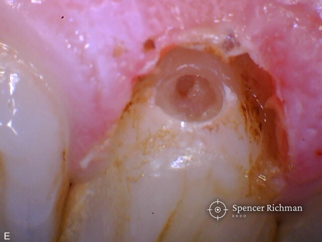
The resorptive cavity exposed at the neck of the tooth, cleaned back to sound tooth structure.
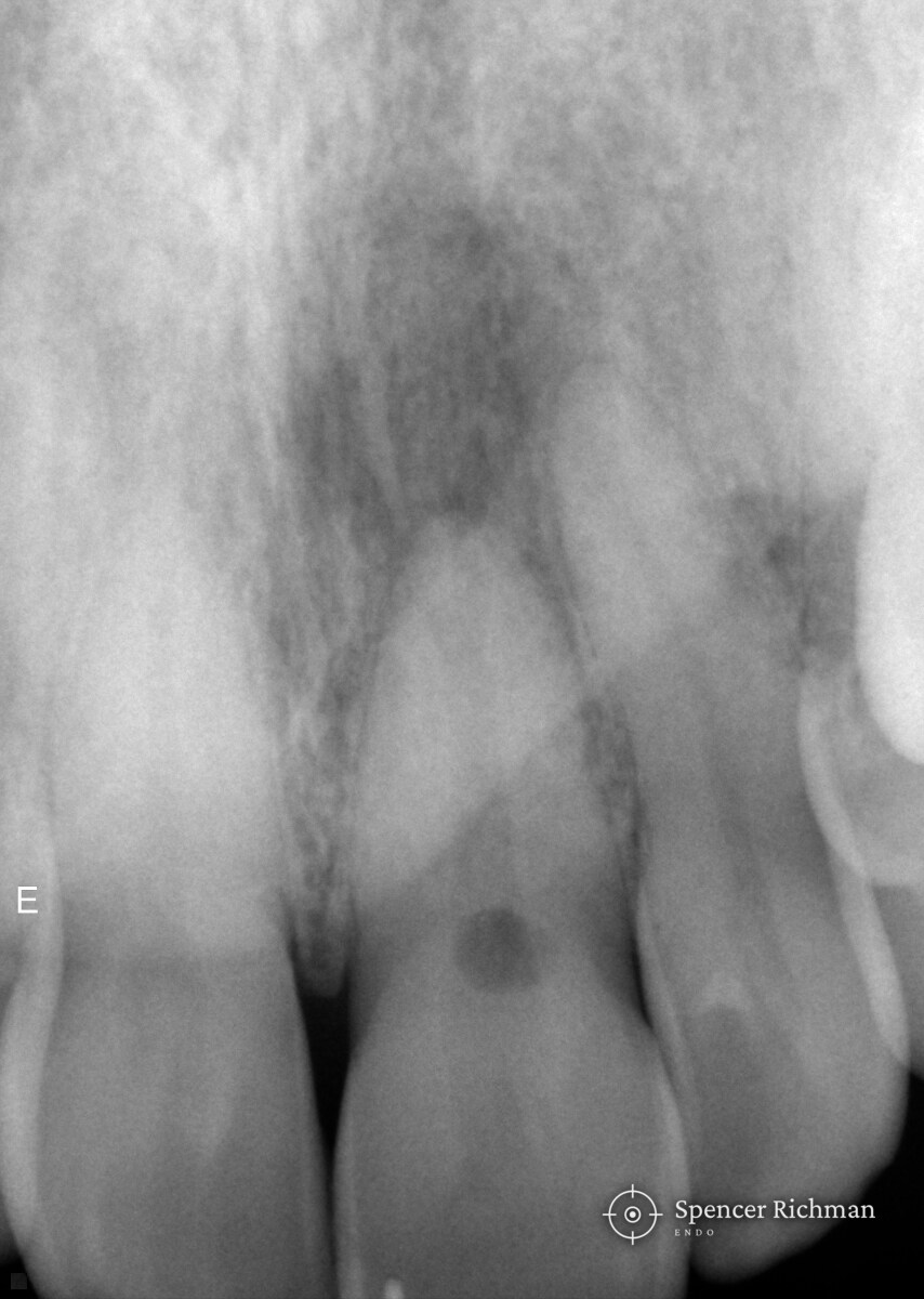
Before treatment
Treatment
Treatment was completed in a single visit at my Woodford Green practice, under rubber dam and the dental operating microscope, with the root canal treatment and the repair both carried out by me at the same appointment.
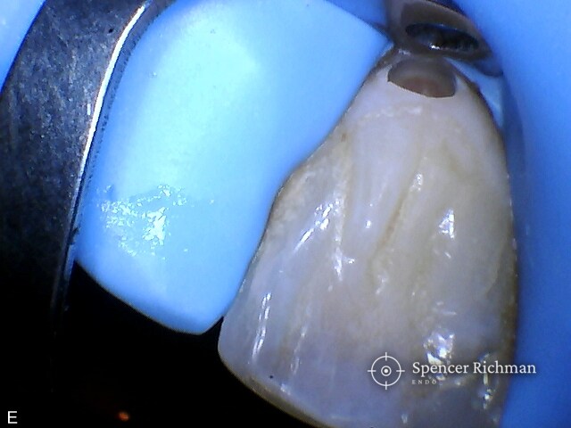
The gum over the defect was recontoured with electrosurgery, which gave clear sight of the resorptive cavity and controlled the bleeding.
A dry field is what the repair depends on, because composite will not bond to a surface that is wet. Rubber dam isolation serves that same end, holding the working area dry and keeping saliva away from the tooth throughout.
The resorptive cavity was cleaned back to sound tooth structure and repaired with composite.
With the defect sealed, the root canal treatment could begin straight away in the same sitting. The canal was shaped with WaveOne Gold, a reciprocating file system.
A tooth treated this way needs watching rather than discharging. This patient was an internal referral, so he continues to be seen within the practice and the tooth will be followed up here.
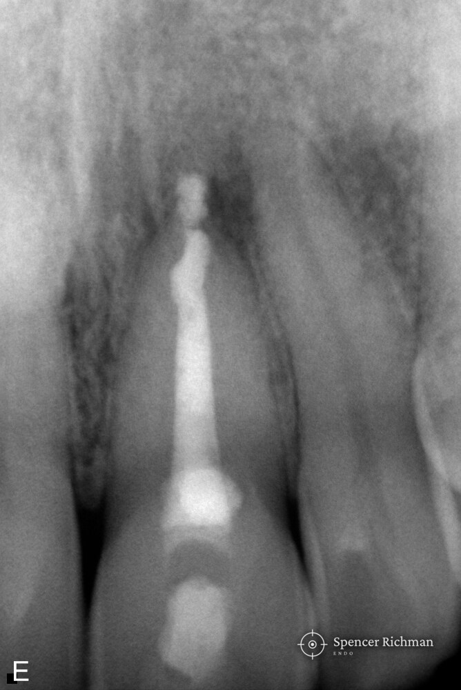
After treatment
Review
Questions patients ask
What is external cervical resorption?
External cervical resorption is a process where the body’s own cells break down the tooth from the outside, starting at the neck of the tooth near the gum line. The process is uncommon, and left alone the damage tends to spread.
Causes, signs and treatment are covered in the guide to external cervical resorption.
Can a tooth injury cause problems years later?
Yes, and a long gap is typical. A knock can damage the surface of the root in a way that only declares years or decades afterwards, by which point most people have forgotten the injury entirely.
Why does resorption cause no pain?
The process is gradual and sits outside the nerve for much of the time. Most people notice nothing until a lump appears in the gum, the tooth changes colour, or a routine X-ray picks the damage up.
What is a lump on the gum above a tooth?
A lump of that kind is often a sinus tract, which is the body’s way of draining infection from inside a tooth. A sinus tract that comes and goes is still a sign of infection, even when nothing hurts.
Can a tooth with resorption be saved?
Often, yes, and how much tooth the resorption has already taken is what decides. Where the defect can be reached and repaired, the tooth can be kept; where the damage runs too deep below the gum, keeping the tooth may not be possible.
Published with the patient’s written consent and fully anonymised. Every tooth is different and outcomes vary from case to case, so this case should not be read as a promise of a particular result.
Get in touch